
What is Mohs Surgery?
Mohs (rhymes with toes) Micrographic Surgery is an advanced treatment procedure for skin cancer which offers the highest potential for recovery – even if the skin cancer has been previously treated. This procedure is a state-of-the-art skin cancer treatment in which the physician serves as surgeon, pathologist, and reconstructive surgeon.
The procedure was developed by Frederic Mohs, M.D. in the 1930’s and has undergone several modifications throughout the years. It relies on the precision and accuracy of a microscope to trace and ensure removal of skin cancer down to its roots. The goal of the procedure is to remove the skin cancer totally while minimizing the amount of normal noncancerous skin removed in the process. The highly trained surgeons that perform Mohs Micrographic Surgery are specialists in both Dermatology and Skin Pathology. Properly trained Mohs surgeons have completed an internship and residency in Dermatology following medical school, plus a fellowship program approved by the American College of Mohs Surgery that includes extensive training in Pathology and reconstructive surgery for at least five years of specialized training beyond medical school.
Click Here to view our Before and After Gallery
The Actual Procedure Is As Follows:
1. A local anesthetic is given at the site of the cancer.
2. A layer of tissue is removed with a small margin around the tumor site. (see Figure 1)
3. A map or drawing is made of the tissue. This is used as a guide to the precise location of any remaining tumor cells.
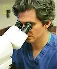
4. The tissue is taken to the lab area in the office and sliced into very thin layers, which are mounted on microscope slides. These are then stained to help see the tissue better (see Figure 2)
5. The entire margin of the tissue is microscopically examined thoroughly to check for evidence of remaining cancer cells.
6. If any cancer is found, the Mohs surgeon returns to the specific area of the tumor site (Figure 3) as indicated on the map and removes another thin layer of tissue only from the area where cancer cells remain. (Figure 4)
7. The newly removed tissue is examined under the microscope.
8. If microscopic analysis still shows evidence of disease, the process continues layer by layer until the cancer is completely gone.
Because this systematic search reveals the complete “roots” of the skin cancer, Mohs surgery offers the highest chance of complete removal of the cancer while sparing normal tissue.
What To Expect If You Have Mohs Surgery?
Do not stop any of your medications including blood thinners unless you are directed to do so by the physician. One week prior to the surgery avoid supplements of vitamin E, Ginkgo, Ginger, Garlic, Ginseng, and Feverfew, which can thin the blood. Three days before and after the surgery, avoid alcohol, which is a mild blood thinner. The day of surgery you may eat your usual breakfast. Please take a shower the morning of the surgery and clean the surgical site and surrounding area well with antibacterial soap and water. Do not wear makeup or apply lotions or creams on or around the area to be treated. Please wear comfortable clothing, preferably a two piece outfit. Due to limited waiting room space, please limit family and friends to one or at most two persons. This will insure your comfort as well as others present for surgery that day. Patients with pacemakers or defibrillators should notify the doctor in advance of their surgery. If you are unable to keep your appointment please notify us within 48 hours and make sure to reschedule your appointment.
Most procedures are completed in 3 to 5 hours. Surgery is performed using a local anesthetic usually at a single visit. Most tumors require 2 to 4 stages for complete removal. There will be about a one hour wait between stages during which each small layer is meticulously examined for remaining cancer cells. You may bring a friend or a loved one to be with you while we are processing the tissue. We also recommend you bring some reading material. After the Mohs procedure has been completed the resulting wound is usually reconstructed to try to achieve the best possible cosmetic and functional results. In certain cases the wound may be allowed to heal on its own. Most patients have only minimal pain after surgery. A normal dose of Tylenol can relieve most discomfort. Some redness or swelling is normal (especially around the eyes) and it gradually decreases in about one week. Bruising goes away in one to two weeks. However, should you experience severe pain or sudden swelling, you should call our office immediately to notify us.
How long has the Mohs procedure been in existence?
The concepts for Mohs micrographic surgery were developed by Frederic E. Mohs, MD, in the 1930s. With the change to the "fresh tissue" techniques and advanced equipment for quickly freezing the tissue in the 1970's, rapid processing of the tissue became more readily available. Since that time, the Mohs micrographic surgical procedure's tissue sparing advantages and high cure rates have been more widely recognized.
How effective is Mohs Surgery?
The cure rate for Mohs Surgery is as high as 99% for basal cell skin cancer and 95% for squamous cell and recurrent cancers. This procedure, the most exact and precise method of tumor removal, minimizes the chance of regrowth and reduces the amount of scarring or disfigurement.
Is Mohs Surgery Always Necessary?
Mohs micrographic surgery is used primarily to treat basal and squamous cell carcinomas, but can also be used to treat less common tumors. Mohs surgery is not typically used to treat melanoma.
Mohs surgery is indicated when:
1. The cancer is in a difficult area where it is important to preserve healthy tissue for maximum functional and cosmetic result such as eyelids, nose, ears, lips, fingers, toes and genitals.
2. Cancers which are large in size (usually greater than 2 cm on the body or extremities and 1 cm on the face.)
3. The edges of the cancer cannot be clearly defined with the naked eye.
4. The cancer grows rapidly or uncontrollably.
5. The cancer exhibits aggressive pathology (related to its appearance under the microscope.)
6. The cancer was treated previously by another method and recurred.
7. Cancers arising in immunosuppressed patients (such as those with a history of organ transplants.)
8. Scar tissue exists in the area of the cancer.
What Is a Mohs Surgeon?
The highly trained surgeons that perform Mohs Micrographic Surgery are specialists in both Dermatology and Skin Pathology. Properly trained Mohs surgeons have completed an internship and residency in Dermatology following medical school, plus a fellowship in a program approved by the Mohs College that includes extensive training in Pathology and reconstructive surgery for at least 5 years of specialized training after medical school.
How Melanoma Is Treated Differently?
Malignant melanoma is typically removed with a wide excision. This involves removing extra skin beyond what can be seen with the eye as melanoma cells can extend well beyond what is visible. The depth reported on the biopsy specimen is important in determining how wide the margins should be when treating the cancer. While there are some centers that remove melanoma with Mohs Micrographic surgery, most do not because it is more difficult to see the abnormal cells on frozen sections used with the Mohs process than with standard processing.